Cholesterosisbulbi
is a condition involving presence of polychromatic, white or golden crystal in
the vitreous cavity and / or anterior chamber. This condition is also known as
hemophthal-mos or synchysis scintillans1. This condition typically
occurs as a sequel of chronic vitreous hemorrhage2 but may occur in
cases of long standing retinal detachment, ocular trauma and advanced Coats
disease3,4. Cholesterol crystals in anterior chamber is a rare
manifestation of this condition, which may be found in advanced cases of
cholesterosis bulbi3,5.
These crystals are
composed of cholesterol which is derived from degradation products of red blood
cells or plasma cells. They can be found freely or engulfed within foreign body
giant cells2. In addition these crystals can also form from
breakdown of vitreous and from subretinal fluid of a long standing retinal
detachment6. In anterior chamber, these crystals can be found in
anterior chamber angle, embedded on iris or may form a hypopyon. In vitreous
cavity, these crystals are found suspended in vitreous which tend to settle
inferiorly when the eye is immobile.
A considerable number of cases with
Cholestero-sisbulbi have been treated with enucleation due to intractable pain
associated with it and the risk of sympathetic ophthalmitis in the other eye.7
We are here to report the first case of Cholesterosisbulbi in Jinnah Hospital
Lahore, associated with profound involvement of anterior chamber with
cholesterol crystals, concurrent increased intraocular pressure and long
standing total retinal detachment.
CASE HISTORY
A 19 year old male was brought to Outdoor
Patient Department of Ophthalmology Unit 1 in Jinnah Hospital Lahore in July
2011 with the primary complaints of painful and blind left eye for last 2
years. His past ocular history revealed surgery on his left eye for congenital
cataract at the age of 4 years (15 years ago) from an eye clinic at Chakwal,
Punjab. Patient was left aphakic after primary surgery. He was prescribed
aphakic spectacles for visual correction. After 3 uneventful years, the patient
suffered from sudden painless and severe decline in visual acuity which converted
to no perception of light after few months of no intervention. He had no
history of trauma and was not using any ocular or systemic medication at the
time of his presentation to us. Family history was also insignificant. The
patient did not have any medical record available for his previous ocular
treatment.
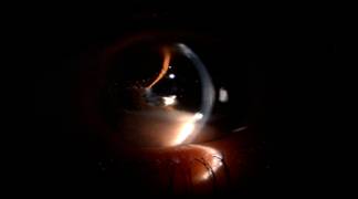
Fig 1: Anterior chamber
photograph showing pseudohypopyon
of polychromatic crystals and shallow anterior chamber.
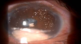
Fig 2: Anterior chamber
photograph showing cholesterol crystals embedded on iris surface.
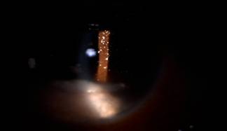
Fig 3: Anterior chamber photograph showing cholesterol crystals
embedded on iris surface.
Fig 4: B-Scan showing total retinal detachment.
Examination of the
eyes showed normal right eye and no perception of light in his left eye. Anterior
segment examination of the left eye showed circum-corneal injection, band
keratopathy, shallow anterior chamber, pseudohypopyon of polychromatic crystals
measuring 3 – 4 mm (Fig 1), polychromatic crystals deposited on iris stroma
(Fig 2, 3), grade 1 flare and +2 cellular anterior chamber activity,
interrupted posterior synechie with non reactive 3mm roughly round pupil and
strongly positive reverse Marcus Gunn reaction, aphakia with intact but
thickened and opacified posterior capsule. Gonioscopy revealed cholesterol
crystals in anterior chamber angle. Goldmann’s tonometry displayed intraocular
pressure of 14 and 32 millimeters of mercury in right and left eye
respectively. There was no view available for examination of vitreous and
retina. B scan ultrasonography showed left sided total retinal detachment (Fig
4).
His rest of general
and systemic examination was unremarkable.
No retinal surgery was advised to the
patient, he was started on topical Atropine 1% three times a day, topical
Dexamethasone 0.1% four times a days, topical Timolol maleate 0.5% two times a
day and oral Acetazolamide 250 mg four times a day. He was also advised
protective polycarbonate glasses for his right eye and to avoid contact sports.
He was asked to follow up after 3 days of initial visit. At his first follow
up, he was found to have intraocular pressure of 27 millimeters of Mercury with
mild reduction in his ocular symptoms. After a month of regular treatment and
follow up and with addition of topical Brimonidine tartrate 0.2% three times a
day, his intraocular pressure was successfully controlled to 18 millimeters of
Mercury with occasional cells in anterior chamber. The patient was also noted
to have significant improvement in his ocular symptom. Currently he is on 15
day follow up with our department.
DISCUSSION
Cholesterol
crystals have been demonstrated in most tissues of eye but the commonest sites
include lens, vitreous and retina. They usually occur as a long term
consequence of ocular trauma, inflammation of uveal tract, degeneration,
particularly of vitreous; and rarely neoplasia7. In a number of
cases, the eye has been blind for a number of years and these crystals have
been found accidently in anterior chamber. Suresh7 conducted
microscopic examination on these crystals and found them to be composed of
cholesterol in the form of thin colorless transparent plates of square or
rectangular shape. Stevens calculated the normal concentration of cholesterol
in normal aqueous and found it to be considerably lower than plasma cholesterol
levels. He also demonstrated the chemical nature of these crystals through
chromatography to be cholesterol8.
The major source of
these cholesterol crystals has been identified to be degenerating red blood
cells either from hyphaema or vitreous hemorrhage9. Long standing
intraocular inflammation resulting in defective blood retinal barrier can also
result in extravasation of cholesterol in the eye and thereafter, its
deposition in different ocular tissues; aphakia is also a recognized cause of
deposition of cholesterol crystals in anterior chamber6. Kennedy6
also reported cases of cholesterosisbulbi involving anterior chamber resulting
after long standing retinal detachments with no evidence of intraocular
hemorrhage as reported in our case. Forsius4 believed that the
process the deposition of cholesterol accelerates when there is clinically
demonstrable evidence of intraocular inflammation because proteins and fats
enter the chamber with the flow of fluid in the eye, and as we know that
cholesterol is insoluble in water, it crystallizes. An important factor in the
deposition of crystals seems to the time for which the eye has remained blind7.
All the seven cases reported by Forsius4 had been blind for more
than 5 years and in Gruber’s series, atleast 6 cases had no sight for more than
5 years. Awan10 reported a case of cholesterol crystals in anterior
chamber of a 15 year old white girl with a structurally and functionally normal
eye.
The cause for high
intraocular pressure can be secondary to deposition of cholesterol crystals in
anterior chamber angle or direct damage of trabecular meshwork by the crystals3.
This was the suspected reason for raised intraocular pressure in our case,
since gonioscopy did not reveal any other angle pathology apart from
cholesterol crystals in angle. Under these circumstances the eye can be made
comfortable by conservative measures as shown by Kumar7. This was
the mainstay of treatment in our patient. In the case reported by Park3,
the causative factor for high intraocular pressure was neovascularization in
anterior chamber angle, which was successfully treated by intravitreal
injection of Bevacizumab along with pars planavitrectomy.
In the past, the mainstay of treatment in
patients with painful blind eyes along with cholesterosisbulbi has been
enucleation, mainly due to ineffective treat-ment available and potential risk
of sympathetic ophthalmitis9,11. With advances in therapeutic
ophthalmology in the form of better anti-glaucoma therapy, potentially more
effective anti inflammatory medications, lasers and anti-vascular endothelial
growth factor agents, a more conservative and cosmetically acceptable approach
has been adopted for such cases. Such cases need to be in a close follow up so
that additional and alternative treatment can be offered in the event of
recurrent or persistently uncomfortable eye.
Author’s affiliation
Dr.
Haroon Tayyab
Medical
Officer
Department
of Ophthalmology
Jinnah Hospital
Lahore
Dr. Muhammad Ali Haider
Medical Officer
Layton Rehmatullah Benevolent Trust
Township, Lahore
Dr. Tehmina Jahangir
Medical Officer
Department of Ophthalmology
Jinnah Hospital
Lahore
Dr. Sana Jahangir
Medical Officer
Department of Ophthalmology
Jinnah Hospital
Lahore
Professor Dr. Samina Jahangir
Head Department of Ophthalmology
Jinnah Hospital Lahore
REFERENCE
1.
Spencer
WH. Ophthalmic
Pathology: An Atlas and Textbook. 4th ed., Philadelphia. Saunders,
1996.
2.
Kanski
JJ, Bowling B. Clinical Ophthalmology: A Systemic
Approach. 7th ed., London. Saunders. 2011; 730.
3.
Park J, Lee H, Kim YK, et al. A Case of Cholesterosis Bulbi with Secondary
Glaucoma Treated by Vitrectomy and Intra-vitreal Bevacizumab. Korean J
Ophthalmol. 2011; 25: 362-5.
4.
Forsius H.
Cholesterol crystals in the anterior chamber. A clinical and chemical
study of 7 cases. ActaOphthalmol (Copenh). 1961; 39: 284-301.
5.
Mielke J, Freudenthaler N, Schlote T, et
al.
Pseudohypopyon of cholesterol crystals occurring 16 years after retinal
detachment in x-linked retinoschisis. KlinMonblAugenheilkd. 2001; 218: 741-3.
6.
Kennedy CJ. The pathogenesis of polychromatic
cholesterol crystals in the anterior chamber. Aust N Z J Ophthalmol. 1996; 24: 267-73.
7.
Kumar S. Cholesterol crystals in the anterior chamber. Br J
Ophthalmol. 1963; 47:
295-9.
8.
Andrews JS, Lynn C, Scobey JW, et al. Cholesterosisbulbi. Case report with modern
chemical identification of the ubiquitous crystals. Br J
Ophthalmol. 1973; 57:
838-44.
9.
Eagle RC Jr, Yanoff M. Cholesterolosis of the anterior chamber. Albrecht
Von Graefes Arch Klin Exp Ophthalmol. 1975; 193: 121-34.
10.
Awan KJ. Crystals in Aqueous Humor of Normal Eye. Ann Ophthalmol. 1978;
10: 37-9.
11.
Yu YS, Kwak HW, Youn DH. Cholesterol crystals in the anterior
chamber. J Korean Ophthalmol Soc. 1980; 21: 117-9.