Superior Limbic
Keratoconjunctivitis (SLK) is a chronic inflammatory condition of the superior
bulbar conjunctiva in a corridor, tunnel or inverted Trapezoid fashion,
associated with the papillary hypertrophy of the upper tarsal conjunctiva. The
first ever description of this condition dates back to the year 1963, when
Theodore and Kimura presented it as a localized, chronic inflammation of the
superior conjunctiva.1 Most common associations of SLK are Thyroid
abnormalities and Dry eye disease. The exact etiology is still unknown but the
final common pathway in this condition is the mechanical soft tissue micro
trauma.2
Since it
is a multifactorial disease, there is no single consensus on the final
treatment. One of the treatment options is topical application of 0.5% to 1%
Silver nitrate. In this particular case report, remarkable results of a single
application of Silver nitrate in SLK, its historical use, application protocol
in SLK, possible mechanism of action, side effects and precautions are
discussed.
CASE
REPORT
A 38 years
old Pakistani female presented in outpatient department with a history of
irritation and redness in both eyes for the last five months. It was associated
with foreign body sensation and watering of both eyes. She had been using
different eye drops but there was no recovery. There was no history of contact
lens use. She was hypertensive, which was not properly controlled. Systemic
history negated any heat intolerance, insomnia and weight loss. However, she
underwent Mastectomy two years back and had been using Tamoxifen since then.
Family history was unremarkable.
On
examination, she was orthophoric and extra ocular movements were of full range.
Pupils were round, regular and normally reacting to light and accommodation.
Visual acuity was 6/6 and intra ocular pressures with Applanation tonometry
were
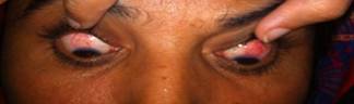
Fig. 1: Superior tunnel shaped congestion in both eyes.
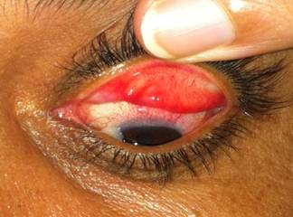
Fig.2: Velvety appearance of the upper tarsal
conjunctiva due to papillary hypertrophy.
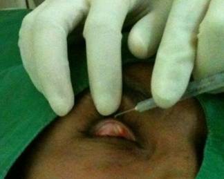
Fig.3: Showing instillation of a drop of 0.5% silver
nitrate on the upper tarsal conjunctiva.
Patient was prescribed topical lubricants and fluorometholone eye
drops. Follow up after three weeks showed mild recovery. We decided to apply
0.5% silver nitrate solution as a trial. Topical Proparacaine was used to
anesthetize the conjunctiva. Upper lid was everted. A drop of 0.5% Silver
nitrate solution was applied to the upper tarsal conjunctiva and the lid was
closed for one minute (Fig. 3). After one minute, the conjunctival sac and cornea
were irrigated with normal saline solution. Slit lamp examination was normal
and Fluorescein staining of the cornea was negative after the procedure.
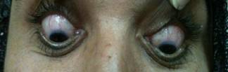
Fig. 4: Marked improvement in superior bulbar congestion.
The patient was asked
to use lubricant eye drops and called for follow up
after three weeks. There was a marked improvement in congestion after three
weeks as shown in (Fig. 4).
DISCUSSION
Superior Limbic
Keratoconjunctivitis (SLK) is a chronic inflammation of the superior bulbar
conjunctiva, distributed in a corridor, tunnel or inverted Trapezoid fashion.
It is associated with the papillary hypertrophy of the upper tarsal
conjunctiva. Theodore coined the term Superior limbic keratoconjunctivitis for
this condition in 1963.1 The exact
etiology of SLK is still unknown but most of the patients
may have abnormal thyroid function3. Studies
have also shown that almost 50% of patients with SLK have
keratoconjunctivitis sicca.4 In this particular patient, Thyroid
function tests and the tear film were normal.
There are certain other risk factors associated with it. These include
prolonged eyelid closure with associated hypoxia, conjunctivochalasis and tight
conjunctival apposition to the globe following upper eyelid procedures.5
How
do all these factors contribute to the superior bulbar congestion, still
remains unsettled. One of the possible mechanisms could be the upper lid
tightness caused by chronic inflammation of the upper bulbar conjunctiva. This
can disturb the normal turnover of the bulbar conjunctival epithelial cells,
which further increases the inflammation.2 Furthermore,
the chronic inflammation can lead to blepharospasm, which presses upon the
bulbar conjunctiva aggravating the existing inflammation.6 Our
patient did not have tight lids but conjunctiva was a bit lax.
There are
certain single case reports available in literature, which increase the
confusion about the etiology of SLK. One case of hyperthyroidism with SLK is
also reported which recovered after resection of the tumor.7 Darrell described
another case of SLK in identical twins proposing a possible genetic basis7. But the family
history of our patient was unremarkable. Whatever be the cause, the final
common pathway in all these conditions is soft tissue trauma.8 It is
hypo-thesized that there are frictional forces between (i) tarsal and bulbar
surfaces; and (ii) between conjunctival stroma and sclera which may be
responsible for this trauma.2
Due to a
multifactorial pathogenesis of SLK, there has been no consensus on a single
best treatment. Various treatment options including artificial tears and
punctal occlusion in dry eyes, alternate patching of the eyes, topical mast
cell stabilizers, vitamin A eye drops, cyclosporin A 0.5%, bandage contact
lenses, cryotherapy and recession or resection of superior bulbar conjunctiva
have all been described in literature with variable success. A case has been reported where unilateral
bandage contact lens has improved the bilateral SLK.9 Thermal and chemical cautery with silver nitrate
has also been used by many clinicians. In one study, a success rate of 73% was
seen with thermal cautery. It was seen that
the number of goblet cells
improved following cautery.10 We decided to try silver
nitrate solution in our patient after many different types of treatments failed
in the previous five months.
Historically,
silver had been mentioned in many literary and medical works since ancient
times. In myths of Vampire stories it was believed that only those bullets
would kill a vampire which contained silver in it. While medical use of silver
salts dates back to 1881 when it was discovered that instillation of a drop of
1% silver nitrate in the eyes of neonates would prevent ocular infections. It
was named Crede prophylaxis after the name of its discoverer.11,12
At that time
it was also called lunar caustic because it was believed by ancient alchemists
that, silver was associated with the moon. It was also used for water storage,
as the water kept in silver containers did not get stale. Some people used to
put silver coins in water utensils as well.
Dramatic
relief of signs and symptoms in our case suggests that the possible mechanism
of action of silver nitrate in SLK is its anti-inflammatory character. The
earliest records of its anti inflammatory action was observed in early 1900
when it was found that if silver nitrate was applied to the indolent wounds,
the inflammation was reduced. In 1920, United States FDA approved silver for
wound treatment.13 Later it was found that Ag ions were released in
water which might have the anti inflammatory action. With the advent of
antibiotics, the use of silver was abandoned and Crede’s prophylaxis became a
history due to corneal burns.
In this
new era of modern medical science there is more research going on Silver nitrate
and not very long ago, it was postulated that the nitrate ions in silver
nitrate had pro inflammatory effect13. This could be the reason that
application of silver nitrate causes irritation and burning as an early effect.
Later this is taken over by the anti-inflammatory effect of silver ions. Slight
burning and irritation noticed in our patient immediately after application of
Silver nitrate could be the result of this effect.
Another
proposed mechanism of action of silver nitrate is its cauterizing effect. There
is 75% silver nitrate with 25% potassium nitrate on a typical applicator of
silver nitrate. As it is applied to a wet surface on the body, nitric acid is
formed. This nitric acid has a chemical cauterizing effect, which is
responsible for the resolution of superior bulbar congestion in SLK. Hence, Silver nitrate, when applied, will achieve its
hemostatic effect by creating chemical cauterization or sealing of the vessels.
We could
not find the results of large prospective studies on the use of silver nitrate
in SLK. However, there are two cases of corneal burns associated with the use
of silver nitrate in SLK where the practitioners had to settle the cases by
large indemnity payments. In one case, Silver nitrate stick was applied to the
tarsal conjunctiva after dipping in Dactriose. The cornea became hazy and final
visual acuity was 20/200. In another
case of a 35 yrs old patient, stick was directly applied to the limbus at 12 O’clock.
A drop of solution dripped on to the cornea causing severe corneal burn at the
spot. It is worth mentioning that in both cases silver nitrate stick was used.14 These sticks are impregnated with concentrated
silver nitrate and should be avoided in eyes. We took special precautions to
avoid these corneal burns. Firstly, the concentration of silver nitrate was
very low. Only 0.5% solution was used. It must be emphasized that if the
required effect is attained after such a low concentration, there is no need to
risk the cornea by using concentrated solutions or sticks. Secondly, the eyelid
was everted to apply the solution to the tarsal conjunctiva rather than
directly applying to the bulbar conjunctiva. Thirdly, the cornea and
conjunctiva were irrigated one minute after the application, to remove excess
of silver nitrate.
So, we make following
recommendations for the use of silver nitrate in SLK.
1.
The surgeon should be
vigilant in using silver nitrate. Solution should not be more concentrated than
1%.
2.
Contact with the skin
should be avoided.
3. The eye should be irrigated after application of the
solution for least 5 to 10 minutes.
4.
Direct contact of silver
nitrate with the cornea should be avoided by everting the lid.
5.
If one has to repeat the
procedure, it should not be before 4 to 6 weeks after the first application.
6.
It is also important that the solution must be kept in a dark and
cool, dry location. If it is not, the medication will degrade and will be
ineffective.
It is always better to use
a freshly prepared solution.
Author’s Affiliation
Dr. Muhammad Khalil
Assistant Professor of Ophthalmolgy
Lahore
Medical and Dental College, Lahore.
Dr. Tayyaba Gul Malik
Assistant Professor of Ophthalmolgy
Lahore
Medical and Dental College, Lahore.
Dr. Sania Munawar
Medical Officer
Ghurki Trust
Teaching Hospital, Lahore
Dr.
Mian Muhammad Shafique
Professor
of Ophthalmology
Lahore
Medical and Dental College, Lahore
REFERENCES
1.
Thygeson
P, Kimura SJ. Chronic conjunctivitis. Trans Am Acad Ophthalmol Otolaryngol. 1963; 67: 494-517.
2.
Cher I. Superior
limbic keratoconjunctivitis: multifactorial mechanical pathogenesis. Clin Experiment Ophthalmol. 2000; 28: 181-4.
3.
Wright P. Superior limbic
keratoconjunctivitis. Trans
Ophthalmol Soc UK. 1972; 92: 555–60.
4.
Udell
IJ, Kenyon KR, Sawa M, Dohlman CH. Treatment of superior limbic
keratoconjunctivitis by thermocauterisation of the superior bulbar conjunctiva.
Ophthalmology 1986;
93:
162.
5.
Sheu MC,
Schoenfield L, Jeng BH. Development of superior limbic
keratoconjunctivitis after upper eyelid blepharoplasty surgery: support for the
mechanical theory of its pathogenesis. Cornea.
2007; 26: 490-2.
6.
Mondino
BJ, Zaidman GW, Salamon SW. Use of pressure patching and soft
contact lens in superior limbic keratoconjunctivitis. Arch
Ophthalmol. 1982; 100: 1932–4.
7.
Roy FW. Fraunfelder FW. Roy and Fraunfelder's
Current Ocular Therapy. 6th edition. P. 393.
8.
Cher I.
Blink-related micro trauma: when the ocular surface harms itself. Clin Experiment Ophthalmol. 2003; 31:
183-90.
9.
Watson S, Tullo AB, Carley F. Treatment of superior limbic keratoconjunctivitis with a
unilateral bandage contact lens. Br J Ophthalmol. 2002; 86: 485-6.
10. Udell
IJ, Kenyon
KR, Sawa
M, Dohlman
CH. Treatment of superior
limbic keratoconjunctivitis by thermo cauterization of the superior bulbar
conjunctiva. Ophthalmology 1986; 93: 162-6.
11.
Grier N. Silver
and its compounds. In: Block SS (ed). Disinfection, Sterilization and
Preservation, Third Edition. Philadelphia, Pa: Lea Febiger; 1983.
12. Peter
H. (2000).
“Dr Carl Credé
(1819 – 1892) and the prevention of ophthalmia neonatorum”. Arch Dis Child Fetal Neonatal. 2000; 83:158–9.
13.
Demling
RH, DeSanti L. Effects of silver on wound management. Wounds.
2001; 13: 4.
14.
Bettman JW. Medication Error Study.
Physician Insurers Association of America, Washington, DC. June 1993. Seven
Hundred Medicolegal Cases in Ophthalmology. Ophthalmology. 1990; 97: 1379-84.