Mundt and Hughes were the first to use ultrasound in ophthalmology
in 19561. They used an (Amplitude) scan for evaluation of
intraocular tumor. Baum and Greenwood introduced the use of B (brightness) scan
in 19582. Both A-scan and B-scan techniques are important for the
diagnosis of ocular diseases3. Diagnostic ultrasound uses sound
waves at frequencies above the range of human hearing (more than 20,000 Hz or
20 KHz)4.
B-scan
ultrasonography is a safe, inexpensive, non-invasive, and accurate tool for
evaluation of the posterior segment of eye when there is media opacity4,5.
It requires the use of high frequency
transducer i.e. a 10 MHz is commonly used for posterior segment assessment.
Nowadays very high-frequency systems (e.g. 50 MHz) can be used for the assessment
of anterior segment of the eye6.
Ultrasonography
has more than 90% sensitivity and specificity in the diagnosis of ocular trauma
cases7. It can detect vitreous hemorrhage, posterior vitreous detachment
(PVD), hemorrhagic choroidal detachment, serous choroidal detachment,
posteriorly dislocated lens, retinal detachment (RD), ocult scleral rupture,
vitreous incarceration and retained intraocular foreign body (IOFB)7.
Bomb blast / mine
blast can cause a variety of potentially blinding posterior segment injuries,
which may be difficult to detect without the use of B-scan ultrasonography. The purpose of our study was to study the role of B-scan
ultrasonography in determining the extent of posterior segment pathology in
blast related eye injuries.
MATERIAL AND METHODS
It was a descriptive case study conducted at Ophthalmology
Department of Khyber Teaching Hospital Peshawar, from March 2010 to February
2012.
Inclusion criteria were unilateral or bilateral eye injury due to
bomb blast / mine blast, both gender & all age groups and poor fundus view
at presentation (due to corneal edema / opacity, hyphema, cataract or vitreous
opacities).
Exclusion criteria were shattered globe in which corneal / scleral
repair was not possible; Consecutive sampling technique was employed i.e. all
the patients who met the inclusion criteria were included in the study.
Detailed
history was taken and complete ocular examination was performed in all cases.
B-scan ultrasonography was done with AB 5500+ A/B Scan (Sonomed, USA) to know
about any posterior segment pathology. In eyes with open globe injury, B-scan
was done after restoring the globe integrity.
RESULTS
Total
number of patients was 97, including 93 males (95.87%) and 4 female (04.12%).
Age of patients was ranging from 4 to 65 years with a mean of 23.70 years.
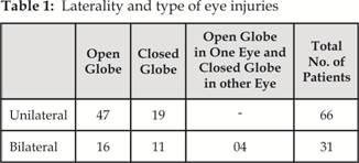
Ocular injury was unilateral in 66 patients (68.04%) and bilateral in 31
patients (31.95%). Of the 128 eyes involved, 83 eyes (64.84%) had open globe
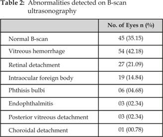
injury and 45 eyes (35.15%) had closed globe injury (Table 1). B- scan was
normal in 45 eyes (35.15%). In the remaining 83 eyes (64.84%) various
abnormalities were detected on B-scan including vitreous hemorrhage, RD, PVD,
choroidal detachment, IOFB, endophthalmitis and phthisis bulbi (Table 2).
Vitreous hemorrhage was the most common pathology seen in 54 eyes (42.18%),
followed by RD which was seen in 27 eyes (21.09%) and IOFB in 19 (14.84%) eyes.
DISCUSSION
Eye injury is a very important cause of visual impairment. Eye
injuries make upto 10% of body injuries, despite the fact that eye makes only
0.27% of the body surface8. Approximately 2 million eye injuries occur in the
United States annually, more than 40 thousand of these results in permanent
visual los9.
Ocular injuries predominantly occur in young males10
and can lead to blindness. Approximately 5% of blindness in the developing
countries is the result of trauma11. Ocular injuries can be divided
into 2 main groups i.e. open globe and closed globe. Open globe eye injuries
include rupture and laceration while closed globe eye injuries include
contusion and lamellar laceration12.
Bomb blasts are a common cause of severe eye injury among adult
males13. They are becoming increasingly common in our country. Bomb blast/ mine
blasts can cause a wide range of potentially blinding posterior segment
injuries14,15.
Direct visualization of the fundus is not possible in eyes with
media opacities such as opaque corneal, hyphema, lenticular or vitreous
opacities16,17. B-scan can help us assess the posterior segment when
the fundus cannot be visualized due to media opacities4.
In this study, 128 eyes with poor view of fundus due to corneal
edema / opacity, hyphema, cataract and/or vitreous opacities were included. One
or more posterior segment pathologies were detected in 83 eyes. Vitreous
hemorrhage was the commonest pathology followed by RD and IOFB.
In one study, ultrasonography detected one or the other pathology
in 21% of ocular trauma cases. Vitreous membrane was seen in 7 %, RD in 6%,
vitreous hemorrhage in 4% & IOFB in 4% cases18. In another study,
ultrasonography revealed RD in 17 (13%), vitreous haemorrhage in 14 (10.7%),
macular edema in 14 (10.7%), endophthalmitis in 12 (9.2%), PVD in 7 (5.4%) and
panophthalmitis in 1 (0.7%) eyes19. Djosevska ED, in his study, detected vitreous hemorrhage 20.9% eyes, RD in 4.4%, endophthalmitis
in 3.3%, PVD in 3.8%, IOFB in 6.6% and choroidal detachment in 1.1% eyes on
ultrasonography20.
In
our study posterior segment pathology was more frequently detected than the
other studies. The reason for this being more severe eye injuries in blast
victims as compared to eye injuries due to other causes. B scan is a very
important diagnostic tool in such patients. The energy used in B-scan ultra-sonography,
does not damage the ocular tissues and it can be repeated (if needed), without
any harmful effects20.
CONCLUSION
B –
scan ultrasonography is a very useful
diagnostic tool for determining the extent of posterior segment pathologies in
blast related eye injuries. In eyes with open globe injury, B – scan
ultrasonography can be safely performed after restoring the globe integrity.
Author’s Affiliation
Dr.
Mumtaz Alam
Assistant
Professor Ophthalmology Department, Kuwait Teaching Hospital Peshawar
Dr.
Akbar Khan
Eye
Surgeon, Khyber Eye Foundation Peshawar
REFERENCES
1.
Mundt GH, Hughes W. Ultrasonics in ocular diagnosis. Am J
Ophthalmol 1956; 41: 488-98.
2.
Baum G, Greenwood I. The application of ultrasonic locating
techniques to ophthalmology, part I: reflective properties. Am J Ophthalmol
1958; 46: 319-29.
3.
Kanski JJ. Imaging Techniques. In:
Kanski JJ Clinical Ophthalmology. A systemic approach 6th ed.
Butterworth Heinemann Elsevier. 2007; 33-58.
4.
Rai P, Shah SIA, Cheema AM, Niazi JH, Sidiqui SJ. Usefulness of B-Scan ultrasonography in ocular trauma. Pak J
Ophthalmol. 2007; 23: 136-43.
5.
Modrzejewska M. The use of ultrasonic
techniques for the diagnosis of retinopathy of prematurity. Ann Acad Med Stetin
2006; 52: 83-8.
6.
Frederic L, Lizzi D, Coleman J.
History of Ophthalmic Ultrasound. J Ultrasound Med. 2004; 23: 1255-66.
7.
Vyas J. Mahesh G. Ultrasonography in Ocular
Trauma. Kerala J Ophthalmol. 2010; 22: 273-6.
8.
Belkin M. A historical
prospective of ocular trauma. In: Miller D, Stegmman R, (edi). Treatment of
anterior segment ocular trauma. Montreal
Medicopia. 1986. p. 7-21.
9.
McGwin G, Xie A, Owsley
C.
The rate of eye injury in the United States. Arch Ophthalmol. 2005; 123: 970-6.
10.
Tielsch JM, Parver LM. Determination
of hospital charges and length of stay for ocular trauma. Ophthalmology. 1990;
97: 231-7.
11.
Thylefors B. Epidemiological
pattern of ocular trauma. Aust NZJ Opthalmol. 1991; 7: 15-8.
12.
Kuhn F, Morris R, Witherspoon CD, Mester V. The Birmingham Eye Trauma Terminology system (BETT). J Fr
Ophtalmol. 2004; 27: 206-10.
13.
Newmann TL, Russo PA. Ocular
sequelae of BB injuries to eye and surrounding adnexa. J Am Optom Assoc. 1998;
69: 583-90.
14. Weichel
ED, Colyer MH, Ludlow SE, Bower KS, Eiseman AS. Combat Ocular Trauma Visual Outcomes
during Operations Iraqi and Enduring Freedom. Ophthalmology. 2008; 115: 2235-45.
15. Rahman
F, Rashid H, Naseem A.
Ocular Sequlae of Blast Injuries:
Experience at a Teaching Hospital. Pak J Med Res 2008; 47: 29-32.
16.
Blumenkanz MS, Byrne SF.
Standardized echography (ultrasonography) for the detection and
characterization of retinal detachment. Ophthalmology. 1982; 89: 821-31.
17.
Rabinowitz R, Yagev R, Shoham A and Lifshitz T. Comparation between clinical and ultrasound findings in patients
with vitreous haemorrhage. Eye 2004; 18: 253-6.
18.
Bhatia IM, Panda A, Dayal Y. Role of ultrasonography
in ocular trauma. Indian J Ophthalmol. 1983; 31: 495-8.
19. Harshadbhai HT, Tyagi M, Jani S, Thakkar J, Sudhalkar A. Paediatric Ocular Trauma and Role of
Echography in Evaluation of
These Cases. AIOC
PROCEEDINGS. Trauma Session. 2010; P. 694-6.
20. Djosevska ED. Ultrasonography in Ocular Trauma. Contri-butions Sec Med Sci. 2013; 34:
105-12.