Original Article
Visual Outcome and Complications of 23 G Versus 20 G Vitrectomy in Cases of Diabetic Vitreous
Haemorrhage
Washoo Mal, Shakir Zafar,
Zafar Iqbal, Syed Fawad Rizvi, Syed Asad Mahmood
Pak J Ophthalmol
2014, Vol. 30 No. 3
. . . .
. . . . . . . . . . . . . . . . . . . . . . . . . . . . . . . . . . . . . . . .
. . . . . . . . . . . . . . . . .. . .. . . . . . . . . . . . . . . . . . . . .
. . . . . . . . . . . . . . . .
See
end of article for authors
affiliations …..……………………….. Correspondence
to: Washoo Mal LRBT Free Base Eye hospital Korangi 2 ½, Karachi Postal code: 74900 E-mail: dr.wash_72@yahoo.com …..……………………….. |
Purpose: To compare post-operative visual outcome and complications of
23-gauge versus 20-gauge pars plana vitrectomy in cases of vitreous
haemorrhage secondary to proliferative diabetic retinopathy. Material and Methods: Randomized clinical trial conducted at LRBT, Free Base Eye
Hospital, Karachi, from January 2010 to June 2012. Two hundred sixty patients
of vitreous haemorrhage secondary to proliferative diabetic retinopathy (Type
– l diabetes mellitus) were randomly selected, age range between 30 – 70
years, 23 – gauge (n = 130) group A; males 73 (56.16%), females 57 (43.85%)
and 20 – gauge pars plana vitrectomy
(n = 130) group B; males 70 (53.85%), females 60 (46.15%). Post-operative
follow up were at day one, 1 week, 1 month, 2 month, 3 month and final 6
month. Data was analyzed and compared for post-operative best corrected
visual acuity (BCVA) and complications between two groups. Results: Visual acuity significantly improved in majority of patients in
both groups. In group A 65.38% (85 patients) achieved between 6/6 - 6/24 and
in group B 63.85% (83 patients) in same range when measured finally at 6 month
post-operative. Post operative complications in group A were transient hypotony 10.77% (14 eyes), recurrent vitreous haemorrhage
6.15% (8 eyes), raised IOP 3.08% (4 eyes), Cataract 3.84% (5 eyes), Itrogenic tear 9.23% (12 eyes), vitreous show (prolapsed)
3.84% (5 eyes) and 1 patient (0.76%) developed endophthalmitis
which was treated successfully. While in group B transient hypotony 6.15% (8 eyes), recurrent vitreous haemorrhage
18.46% (24 eyes), raised IOP 7.69% (10 eyes), Cataract 3.84% (5 eyes), Itrogenic tear 7.69% (10
eyes) and phthisis bulbus 0.76% (1 eye),
corneal edema 6.92% (9 eyes), and conjunctival
granuloma at surgical wound 2.30% (3 eyes) observed. Conclusion: 23 – gauge micro-incision Vitrectomy
system MIVS and 20-gauge pars plana vitrectomy showed improvement in best
corrected visual acuity (BCVA) while early visual recovery and less
complications rate seen in patients of 23 – gauge group. Key words: Vitrectomy, Diabetic
retinopathy, Vitreous haemorrhage. |
It has
been estimated that 8.5 – 12% population Pakistan suferring
from diabetes mellitus, type-ll is more common and prevalence
of proliferative diabetic retinopathy 2.65 – 5% in our country.1,2 Vitreous haemorrhage due to proliferative diabetic
retinopathy (PDR) is one of major cause of visual impairment and due to
emergence of vitrectomy system and rapid advancement
in surgical techniques for posterior segment pathologies led to improvement in
visual outcome after surgery.
Pars plana vitrectomy (PPV) is one of
the most commonly performed surgical procedure for treatment of various vitreo-retinal diseases. Machemer
et al introduced PPV in 19713. More than Last 30 years, the 3-port
20-gauge PPV remained the standard vitreo-retinal
surgery4. Pars plana vitrectomy involves conjunctival
incision, sclerotomies and suturing but now-a-day’s
20-gauge PPV is also performed transconjunctivally to
make entry wound through the conjunctiva and sclera together, which does not
required conventional suturing. These sutureless self sealing sclerotomies for PPV were first designed by Chen in 19965.
In
2002, Fujil et al introduced the 25-gauge transconjunctival sutureless micro-incision vitrectomy surgery (MIVS).6,7
After few years, in 2005 Eckardt developed the
23-gauge transconjunctival vitrectomy system8.
Though the advances in surgical equipments and techniques,
the vitrectomy procedure requires a skilled and experienced surgeon.
Conventional 3-port 20-gauge PPV need 1.2 mm wide sclerotomies,
performed after conjunctival peritomy
and surgical wound secured with sutures that may cause sub-conjunctival
haemorrhage, congestion and foreign body sensation responsible for
post-operative discomfort as well as prolong surgical time9. In
recent years, the 23-gauge transconjunctival
sutureless vitrectomy (TSV) has become the preferred MIVS system of vitreo-retinal surgeons because of its smaller sclerotomies of 0.72 mm width and leads decrease surgical
trauma, minimum post-operative inflamma-ion, faster
healing and due to its sutureless, shortens the surgical time.10-12
The 23-gauge vitrectomy system has advantage to
overcome the excessive flexibility of instruments used in smaller diameter
25-gauge MIVS, which may cause hindrance in maneuvering the globe during
surgery13.
The
rationale of this study is to compare post-operative visual outcome and
complications between 23–gauge MIVS and 20–gauge pars plana
Vitrectomy.
MATERIAL AND METHODS
In this prospective, randomized clinical trial of 260 patients of
vitreous haemorrhage secondary to proli-ferative
diabetic retinopathy (Fig. 1) were randomly selected for 23 – gauge MIVS (n = 130)
and 20 – gauge PPV (n = 130) for indication of diabetic vitreous haemorrhage
during period of January, 2010 to June, 2012, carried out in LRBT, Free Base
Eye Hospital, Karachi. All surgeries were carried out by one vitreo-retinal surgeon (SZ). The data acquisition was
performed by two investigators (ZI) independently of surgeon, while literature
was reviewed by doctor (WM). The study was conducted under the supervision of
hospital incharge (SFR). The study was approved by
institutional ethical review committee, and informed consent was taken from
each patient.
Cases of diabetic vitreous haemorrhage secondary to type – II
diabetes mellitus, without significant cataract and patients with pseudophakia were included in this study while patients associated
with advanced or complicated PDR like retinal detachment, proliferative vitreo-retinopathy, uveitis, dense cataract and previous
history of vitreo-retinal surgery were excluded from
study and in invisible fundi dynamic B-scan ultrasound carried out to rule out
retinal detachment or any significant fibrotic bands. Pre-operative ocular
& systemic examination of all patients done, including: visual acuity (Snellen’s Chart), ocular adnexa, anterior segment
examination, intra ocular pressure, crystalline lens for opacities (cataract)
and dilated fundal examination with +90 D lens at bio-microscopic slit lamp,
indirect ophthalmoscopy, blood pressure, fasting blood sugar and HbA1c were
checked. All patients were informed about the procedure and written consent
taken. Surgeries were performed by using vitrectomy system) and non-contact
viewing system (EBIOS) for visualization and illumination provided by Xenon
light.
The
rationale of this study is to compare post-operative visual outcome and
complications between 23-gauge MIVS and 20-gauge pars plana
vitrectomy.
Surgical Technique
All
surgeries done under local anaesthesia preferably retrobulbar
with 2 ml of lidocain 2% and 2 ml of bupivacain 0.75% were used. All surgeries were carried out
under strict aseptic measures using povidone iodine
5-10% for periocular paint and conjunctival
fornices with 5% same solution. After applying eye
speculum, self-retaining trocar/cannulae inserted transconjunctivally after 1.5-2 mm displacing conjunctiva
laterally at supero-temporal, supero-nasal
and infero-temporally about 3.5 mm away from the limbus, 300 obliquely to have sclera tunnel in
all phakic patients. The infusion line was connected
to infero-temporal cannula while superior two cannulae for illumination and vitrectomy cutter, after
complete vitreous cleaning, the retina was examined and pan retinal
photocoagulation was done using endolaser (Fig: 2).
Balance salt solution was used as internal tamponade.
At the completion of vitrectomy the superior cannulae were plugged and infusion line was stopped.
Initially superior cannulae were removed with
observing the repositioning of conjunctiva covering the sclerotomies
finally infero-temporal cannula with infusion line
removed and observed repositioning of conjunctiva; finally the sclerotomies in group A, of 23-gauge MIVS left sutureless. (No any patient required suturing in group AIn Group B, 20-gauge sclerotomies
were done with MVR blade and infero-temporally
infusion canula was sutured with vicryl
6/0. At end of surgery, the sclerotomies and
conjunctiva were sutured with vicryl 6/0 (Ethicon,
Johnson and Johnson).
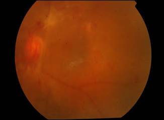
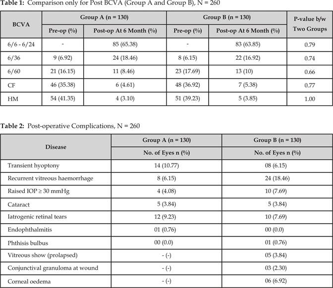
Fig. 1:
Pre operative Fundus photograph of Vitreous Haemorrhage
Fig. 2: Fundus photograph after Pars Plana Vitrectomy
with Endolaser
The data was collected for
variables like, age, gender, best corrected visual
acuity (BCVA). Pre-operatively and post-operatively follow up were at day one,
1 week, 1 month, 2 month, 3 month and finally at the end of 6 months visual
acuity measured with Snellen’s chart followed by
refraction (where needed) and various post-operative complications were
evaluated. Variables were statistically analyzed by Wilcoxon test for pre &
post operative BCVA and Chi-square’s Test and Fisher’s Exact Test where applied
for comparison two groups. A P-value
≤ 0.05 considered statistically significant.
RESULTS
Data of
two hundred sixty eyes of 260 patients were analyzed, 130 in each group A for
23-gauge MIVS and group B for 20-gauge PPV. Age range was 30 to 70 years, 73
(56.15%) males and 57 (43.85%) females in group A while 70 (53.85%) males and
60 (46.15%) females in group B. BCVA of two groups were analysed
by applying Wilcoxon test (NPar) to compare pre and
post BCVA of two groups, that showed significant improvement (p-value 0.0001).
BCVA differences in patients of
two groups were insignificant when measured finally at 6 months
post-operatively shown in Table 1. In group A 85 (65.38%) improved between 6/6
- 6/24, 6/36 24 (18.46%), 6/60 11 (8.46%), CF 6 (4.61%), HM 4 (3.10%); whereas
group B improved 83 (63.85%) - 6/6 - 6/24, 22 (16.92%) - 6/36, 13 (10%) - 6/60,
7 (5.38%) - CF, 5 (3.85%) -HM (HM due to ischemic maulopathy
proven by FFA and recurrent vitreous haem:). P-value
of BCVA between two groups remained insignificant. Early visual recovery observed
in group A that might be due to lesser manipulation.
Post-operative complications are summarized the table 2. Only one patient
(0.76%) in group A developed endophthalmitis which
was successfully treated with standard intra-vitreal,
topical and systemic antibiotics and one eye (0.76%) in group B end up into
phthisis bulbus. Inspite of
itrogenic tears, no any patient developed retinal
detachment. Because confluent lasers were applied around
tears.
DISCUSSION
In this
study post operatively BCVA of both groups at 6 months significantly improved
shown in (table 1) from hand movement to between 6/6 to 6/24 in 65.38% (85/130
patients) in 23-gauge group and 63.85% (83/130 patients) in 20-gauge group;
which shows insignificant statistical difference between two groups. A study of
Kim JM et al shows BCVA 6/6 to 6/24 in 72.72% in cases of vitreous haemorrhage
23-
gauge vitrectomy which is much better
improvement than this study14. This difference in visual outcome is
due to variation in case selection. Nataraj AMS mentioned
significant BCVA improvement in his study of 23-gauge and 20-gauge technique
with insignificant statistical difference in two groups, which is comparable to
this study15.
Complications
summarized in table 2, showing hypotony (defined as ≤
5 mm Hg intraocular pressure)16 occurred in
10.77% (14/130 eyes) in 23-gauge and 6.15% (8/130 eyes) in 20 – gauge groups
which was normalized in 2 weeks with use of topical steroids and cycloplegics. Woo SJ et al noted post-operative hypotony in 11.3% in 23-gauge vitrectomy17 and
these results are comparable to this study. Related to this multiple studies
and literature shows increase incidence of post-operative hypotony
in sutureless vitrectomy.6,11,18
Intraocular pressure ≥ 30 mm Hg was observed in 3.08% (4/130 eyes) in
23-gauge and 7.69% (10/130 eyes) in 20 – gauge groups. This would be due to
suturing of sclerotomies in respective groups. Misra A et al mentioned raise IOP
8% (4/50 eyes) in 20-gauge vitrectomy, that incidence is matched to this study.
Iatrogenic retinal breaks in 23-gauge and 20-gauge groups are 10.77% (14/130
eyes) and 9.23% (12/130 eyes) respectively. Misra A et al also shows iatrogenic breaks 14% and 12% in 23 – gauge
and 20 – gauge respectively which is higher than this study19.
As the
23-gauge vitrectomy is sutureless nature of surgery
is vulnerable to most serious complication - the endophthalmitis,
Kunimote et al in one large retrospective case series
has shows incidence of endophthalmitis after
sutureless 25 gauge (MIVS) is 12 times higher than conventional 20-gauge
vitrectomy20. However in subsequent case series of Hu AY et al and Parolini B et al have shows no increase incidence of endophthalmitis in 25 – gauge and 23-gauge compared with
20-gauge vitrectomy21,22. In this study single
case developed endophthalmitis, this was successfully
treated with intravitreal, topical and systemic antibiotics. The cause of post
surgical endophthalmitis might have involved factors
other than sutureless vitrectomy. Misra A et al and Romano MR mentioned in
their studies that sutureless vitrectomy theoretically may be at greater risk
of this dangerous complication.19,23
Recurrent vitreous hemorrhage
noticed in 8% (21 cases in both groups) more probably due to underlying basic
pathology of proliferative diabetic retinopathy.
CONCLUSION
This study concludes that 23 – gauge
MIVS and 20 – gauge pars plana vitrectomy
showed improvement in best corrected visual acuity (BCVA), while low rate of
complications seen in 23 – gauge MIVS except hypotony
is higher due to sutureless surgery and preferably MIVS should be considered
first choice where ever possible.
Author’s Affiliation
Dr. Washoo Mal
Associate
Ophthalomologist
LRBT
Free Base Eye Hospital
Korangi 2 ½, Karachi
Postal
code: 74900
Dr. Shakir Zafar
Consultant
Ophthalmologist
LRBT
Free Base Eye Hospital
Korangi 2 ½, Karachi
Postal
code: 74900
Dr. Zafar Iqbal
Consultant
Ophthalmologist
LRBT
Free Base Eye hospital
Korangi 2 ½, Karachi
Postal
code: 74900
Dr. Syed Fawad Rizvi
Chief
Consultant Ophthalmologist
LRBT
Free Base Eye Hospital
Korangi 2 ½, Karachi
Postal code: 74900
Dr.
Syed Asad Mahmood
Resident
medical officer
LRBT
Free Base Eye hospital
Korangi 2 ½, Karachi
Postal
code: 74900
REFERENCES
1.
Memon WU, Jadoon Z, Qidwai U, Naz S, Dawar S, Hasan T. Prevalence of
diabetic retinopathy in patients of age group 30 years and above attending
multicentre diabetic clinics in Karachi. Pak
J Ophthalmol. 2012; 28: 99-104.
2.
Hussain F, Arif M,
Ahmad M. The prevalence of
diabetic retinopathy in Faisalabad, Pakistan: a population-based study. Turk J
Med Sci. 2011; 41: 735-42.
3.
Machemer R, Buettner H, Norton EW, Parel JM.
Vitrectomy: a pars plana approach. Trans Am Acad Ophthalmol Otolaryngol. 1971; 75:
813-20.
4.
O’Malley
C, Heintz RM Sr. Vitrectomy with an
alternative instrument system. Ann Ophthalmol. 1975;
7: 585-8.
5.
Chen JC.
Sutureless pars plana vitrectomy through self-sealing sclerotomies.
Arch Ophthalmol. 1996; 114: 1273-5.
6.
Fujii GY, De
Juan E Jr
, Humayun MS, Pieramici
DJ, Chang TS, Awh C, Ng E, Barnes A, Wu SL, Sommerville DN. A new 25-gauge instrument
system for transconjunctival sutureless vitrectomy
surgery. Ophthalmology. 2002; 109: 1814-20.
7.
Fujii GY, De
Juan E Jr, Humayun MS,
Chang TS, Pieramici DJ, Barnes A, Kent D. Initial
experience using the transconjunctival sutureless
vitrectomy system for vitreoretinal surgery.
Ophthalmology. 2002; 109: 1814-20.
8.
Eckardt C. Transconjunctival sutureless 23-gauge vitrectomy. Retina.
2005; 25: 208-11.
9.
Jackson
T. Modified sutureless sclerotomies in
pars plana vitrectomy. Am J Ophthalmol. 2000, 129:
116-7.
10. Meyer CH, Rodrigues EB, Schmidt JC, horle S, Kroll P. Sutureless vitrectomy surgery.
Ophthalmology. 2003; 110: 2427-8.
11. Soni M, ChHugh D.
23-Gauge transconjunctival sutureless vitrectomy: a
way forward. Eye News. 2007; 14: 18-20.
12. Lakhanpal RR, Humayun MS.
De juan E Jr, Lim JI, Chong
LP, Chang TS, Javaheri M, Fujii GY, Barnes AC,
Alexandrou TJ. Outcomes
of 140 consecutive cases of 25-gauge transconjunctival
surgery for posterior segment disease. Ophthalmology. 2005; 112: 817-24.
13. Nam Y, Chung H, Lee JY, Kim JG, Yoon YH.
Comparison of 25 and 23-gauge sutureless micronicision
vitrectomy surgery in the treatment of various vitreoretinal
diseases. Eye 2010; 24: 869-74.
14. Kim JM, Park HK, Hwang MJ, Yu GH, Suk Y,
Yu, Chung H. The safety and efficacy of transconjunctival
sutureless 23-gauge vitrectomy. Korean Journal of Ophthalmology. 2007; 21:
201-7.
15. Nataraj AMS. Comparison Between 20 – gauge
and 23-gauge Vitrectomy In
Diabetic Patients. Kerala Journal of Ophthalmology. 2011; 23: 293-7.
16. Kanski JJ, Bowling B. editors. Acquired
macular disorders. In: Clinical ophthalmology a systemic approach. Edinburg:
Elsevier; 2011;.594-646.
17. Woo Sj, Park KH,
Hwang JM, Kim JH,
Yu YS, Chung H.
Risk factors associated with sclerotomy
leakage and postoperative hypotony after 23-gauge transconjunctival sutureless vitrectomy. Retina. 2009; 29: 456-63.
18.
Fine HF,
Iranmanesh R, Iturralde D, Spaide RF. Outcomes of 77 consecutive
cases of 23-gauge transconjunctival vitrectomy
surgery for posterior segment disease. Ophthalmology. 2007; 114: 1197-1200.
19.
Misra A,
Ho-Yen G; Burton RL. 23-gauge Suturless Vitrectomy and
20-gauge Vitrectomy: A Case Series Comparison. Eye. 2009; 23:1187-91.
20.
Kunimoto DY,
Kaiser RS. Wills Eye Retina Service. Incidence of endophthalmitis
after 20 and 25-gauge vitrectomy. Ophthalmology. 2007; 114: 2133-7.
21.
Hu AY,
Bourges JL, Shah SP, Gupta A,
Gonzales CR,
Oliver SC,
Schwartz SD. Endophthalmitis after pars plana vitrectomy a 20 and 25 – gauge
comparison. Ophthalmology. 2009; 116: 1360-5.
22.
Parolini B, Romanelli F, Prigione G, Pertile G. Incidence of endophthalmitis in a large series of 23 – gauge and
20-gauge transconjunctival pars plana vitrectomy. Greafes Arch Clin Exp
Ophthalmology. 2009: 247: 1711-2.
23.
Romano
MR, Das R, Groenwald C, Stapler T, Marticorena J, Valldeperas X,
Wong D, Heimann H. Primary 23-gauge
sutureless vitrectomy for rhegmatogenous retinal
detachment. Indian J Ophthalmol. 2012; 60: 29-33.