Loa loa is a parasitic infection endemic in the tropical rain
forests of Africa. It is unique among the human filarial Infestation and adult
worms are occasionally visible during subconjunctival
migration. This case report is the removal of a dead Loa Loa
worm from subconjunctival space of a patient who came
in the ophthalmology department of Al-Khidmat
Teaching Hospital Mansoora. Medline search revealed
this as the first case in
CASE REPORT
We report a case of
removal of sub conjunctival Loa Loa
of a patient aged 70, who belongs to Phool Nagar (Bhai Pheru), about 40 Km from
(Fig. 2). One end of the worm was curled up. It was dissected out and measured
about 2.8 cm in length (Fig. 3). We suspected worm to be Loa Loa and sent it for parasitological examination. The
patient did not give any history of travel to Africa except a visit to
DISCUSSION
Loa loa belongs to super family Filarioidea.
Adult worm is long thread like. They are parasites of subcutaneous tissues or
serous cavities as sub conjunctival spaces. The worms
are viviparous that they give birth to larvae and do not lay eggs. They
commonly migrate rapidly in the body and may be seen in sub conjunctival
space or thinned skin areas. Adult worm measures 3 cm in length and 350 micron
meter in width. Female worm measures 6 cm in length and 450 micron meter in
breadth. Its vector, in which the parasite undergoes larval stages, is a blood
sucking fly of the genus chrysops.1 There
are some reports of worm located in the anterior chamber of the eye.2
The worm
causes Loais is characterized by the occurrence of
swelling in various parts of the body known as calabar
swellings. The swellings are transient and may be painful if situated over
joints. They are caused by maturing larvae migrating away from the site of
inoculation by vector fly. Eosinophilia is common.

Fig.
1: Patient
of subconjunctival Loa Loa
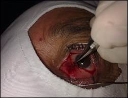
Fig.
2:
During Surgery
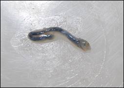
Fig.
3:
Loa Loa Worm after Removal
Most of
the cases of sub conjunctival Loa Loa
reported are live worms while one case report of dead worm recovered from eye
in Brazil 3-5. History of travel to Africa is usual in most of the
case reports.6-8 The disease was also found
in some of the African students studying abroad9. The worm also
recovered from periocular subcutaneous tissue in few
reports.10,11
We report a case
with subconjunctival Loaiasis.
In summary, this is a case of Loaiasis encountered in
non-endemic area. There is no history of travel to
Author’s Affiliation
Dr.
Mohammad Mateen Amir
Associate
Professor of Ophthalmology
University
of Lahore & Head of Department
Al- Khidmat Teaching Hospital Mansoora
Lahore
Dr.
Afsar Saeed Shaikh
Associate
Professor Pathology
Dr.
Ameena Ashraf
Associate
Professor Pathology
REFERENCES
1.
Roberts
LS, John Janoy JR: Foundation of parasitology. 8th ed; McGraw Hill International.
2010; 472.
2.
Barua P, Barua N, Hazarika NK, Das S. Loa Loa in the anterior chamber of the eye: a case report.
Indian journal Medical Microbiology. 2005; 23: 59-60.
3.
Carme B, Botaka E, Lehenaff YM. Dead LoaLoa
filarial in a subconjunctival site. Apropos of a
case. Journal French Ophthalmol. 1988; 11: 865-7.
4.
Bowler GS, Shah AN, Bye LA, Saldana M.
Ocular Loiasis in London 2008-2009: A case series.
Eye (
5.
Carbonez G, Oogziekten. Lindendreef 1, 2020 Antwerpen. Subconjunctival LoaLoa worm: Case
report. Bull Soc Ophthalmol,
2002; (283): 45-8.
6.
Aiello F,
Palma S, Varesi C, Cerulli
A, Valente R, Ajello
L.
A rare case report of Loa Loa ocular filariasis. Europeon Journal Ophthalmol. 2010; 20: 237-9.
7.
Eballe AO, Epee E, Koki G, Owono
D, Myogo CE, Bella AL. Intraocular live
male filarial Loa Loa worm. Clinical ophthalmol. 2008. Dec; 2. 965-7.
8.
Wickremesinghe RS,
9.
Ali S,
Fisher M, Juckett G. The African eye
worm: A case report and review. J Travel Med. 2008; 15: 50-2.
10.
Sbeity ZH, Jaksche A,
Martin S, Loeffler KU. Loa Loa Microfilariasis in the
eyelid: case report of the first periocular
subcutaneous manifestation in
11.
Bhedasqaonkar S, Baile RB, Nadkarni S, Jakkula G, Gogri p. Loa Loa macrofilaiasis in the eyelid: Case report of the first periocular subcutaneous manifestation in