Xeroderma Pigmentosum
(XP), first described by Hebra and Kaposi in 1874,1
is a rare autosomal recessive genetic disorder. It is characterized by faulty
repair of DNA damage induced by ultraviolet radiation. XP occurs worldwide,
affecting all age groups, both sexes and all racial groups. The basic
deficiency lies in the nucleotide excision repair (NER), a mechanism
responsible for recognizing and repairing bulky DNA damage caused by
environmental and other exposures, thus resulting in the clinical
manifestations.
Fibroblasts
in normal human skin can repair damage caused by exposure to UV radiation.
However, in patients with XP, this ability of fibroblasts is slower or
completely absent.2 XP have many
consequences including skin cancer, ocular abnormalities (e.g., conjunctivitis,
ectropion and corneal opacities) and neurological
anomalies resulting in decreased reflexes, progressive hearing loss and mental
retardation.3 Here, we describe
four cases of this disease in a single Pakistani family.
OUR XP FAMILY
An 11
year old girl, resident of Karachi, Pakistan presented to us with complaints of
loss of vision in her right eye, intolerance to light and mild pain and ocular irritation
for the past 4 years. The symptoms had increased in the last 4 months. Her left
vision was completely lost 3 years back. She had a strong family history of XP.
Four out of her 7 siblings suffered from it. Two elder siblings, one elder
brother and one elder sister, died because of XP – related complications at
ages 18 and 9 respectively. Their parents were first cousins. On examination
multiple hypo and hyper pigmented areas of skin around the eyes were visible.
Similar lesions were observed on the scalp with loss of hair. There was no
evidence of systemic malignancy. Her visual acuity in the right eye was hand
movement. Her left eye was phthysical with no
perception of light. Examination showed right eye madarosis,
completely opaque and dry cornea with
peripheral corneal vascularization. Based on her signs and symptoms and
the associated family history, a clinical diagnosis of xeroderma
pigmentosum was confirmed.
DISCUSSION
We reported a single
Pakistani family with 4 of 7 siblings affected by severe xeroderma
pigmentosum. Two of them died at the age of 9 and 18
years, respectively, while the two survived with unilateral blindness and
extreme sensitivity of the skin.
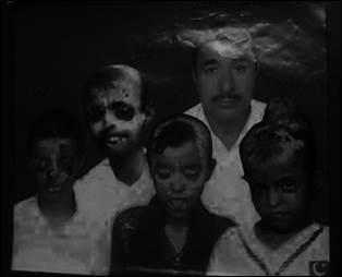
Fig. 1: A picture of the family including the patient and her siblings
affected by XP
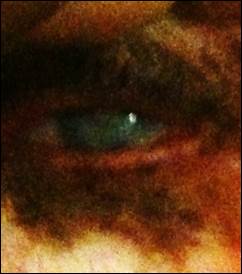
Fig. 2: Picture taken while examining the patients’ eye (left or right).
Visible pigmentation of skin near the eye
There are multiple
manifestations of XP including cutaneous, ocular and neurological - some more prevalent
and severe than others. For example, among 36 cases studied by Bhutto and
colleagues(18 males and 18 females, age range 2 - 30 years)4
in the dermatology unit of a
tertiary care hospital in Larkana or medical camps in remote areas over a period
of seven years, two thirds had severe disease. They also found that 29 (81%)
cases had ocular symptoms including photophobia, conjunctivitis, corneal
keratitis and lid ulcer. One patient had complete bilateral loss of vision.
Both the family members we
examined had unilateral loss of vision and severe ocular damage. It is
well-established that ocular changes are more common in the tissues exposed to
UV light, such as the eyelids, conjunctiva, cornea, and the lens.5
Photophobia is often the
first symptom to appear followed by pigmentation of eyelids, madarosis, ectropion and lower
lid cancer. Conjunctival damage results in xerosis, telangiectasia, chronic congestion, and pigmentry changes whereas involvement of the cornea results
in dryness, exposure keratitis, hazyness, band-like
nodular keratopathy and scarring and ulceration,
resulting in severe visual impairment. Other reasons for visual loss in XP
patients could be pterygium, tumour
invasion from the limbus, and corneal
vascularization.6
The diagnosis of XP in this
case series was based on clinical findings and positive family history.
Unfortunately, diagnostic tests were not available in our setting.
Although there is no cure for
XP, an important measure of protection from sunlight is adopted to overcome
skin damage. This is carried out by covering windows with UV resistant films
and application of sun screen on exposed skin. Since avoiding sunlight may
result in Vitamin D deficiency, it is advisable to prescribe Vitamin D supplements.
Other protective actions like frequent eye examinations and removal of
pre-cancerous lesions are also advised. More importantly the patient is offered
psychological support to improve his quality of life. The common problems that
need to be addressed here are feelings of isolation and career prospects. There
are XP support groups in developed countries such as France, Germany, UK and
USA. They offer a wealth of advice and help. Unfortunately, such groups do not
exist in our part of the world and need to be established. Moreover, genetic
counseling and testing is also an important component of its prevention which
is not readily available in Pakistan.
In conclusion, the four cases of XP we reported
had devastating ocular consequences.
Author’s Affiliation
Dr. Najia Idrees
Section
of Ophthalmology
Department
of Surgery
Aga
Khan University
Karachi
Dr. Tanveer Anjum Chaudhry
Section
of Ophthalmology
Department
of Surgery
Aga
Khan University
Karachi
Dr. Arsalan Rajput
Section
of Ophthalmology
Department
of Surgery
Aga
Khan University
Karachi
REFERENCES
1.
Grampurohit VU, Dinesh US, Rao R. Multiple
cutaneous malignancies in a patient of xeroderma pigmentosum. Journal of cancer
research and therapeutics. 2011; 7: 205-7.
2.
Cleaver JE. Defective repair replication of
DNA in xeroderma pigmentosum. Nature. 1968; 218: 652-6.
3.
Kraemer KH, Lee MM, Scotto J.
Xeroderma pigmentosum. Cutaneous, ocular, and neurologic abnormalities in 830
published cases. Archives of dermatology. 1987; 123: 241-50.
4.
Bhutto AM, Shaikh A, Nonaka S.
Incidence of xeroderma pigmentosum in Larkana, Pakistan: a 7-year study. The
Br. J of dermatology. 2005; 152: 545-51.
5.
Lehmann AR, McGibbon D, Stefanini M.
Xeroderma pigmentosum. Orphanet journal of rare diseases. 2011; 6: 70.
6.
Goyal JL, Rao VA, Srinivasan
R, Agrawal K. Oculocutaneous manifestations in xeroderma
pigmentosa. The Br. J of Ophthalmology. 1994; 78:
295-7.