Vision 2020 is the global initiative, launched in 1999 by the
International Agency for the Prevention of Blindness (IAPB) and World Health
Organization (WHO), with the aim of eliminating avoidable blindness. In
Pakistan, the national survey done in 2006 showed prevalence of blindness to be
3.4% and severe visual impairment as 4.9% in patients who were 30 years or
older1. Significant development has been noted in treatment and
prevention options of anterior segment eye diseases like cataract and trachoma but
a large proportion of avoidable blindness in developing countries of Asia is
due to posterior segment diseases such as glaucoma and diabetic retinopathy2.
Pakistan has 6th largest population in the world. Diabetic Association
of Pakistan (DAP) and WHO showed an overall prevalence of diabetes as 11.47%
(ranged from 6.39–16.5%)3. According to internal diabetic federation
(IDF), there were 6.9 million cases of diabetes in Pakistan in 2014 and
prevalence of diabetes in adults of 20-79 years of age was 6.8%. However, the
projected estimates of International Diabetic foundation (IDF) for 2035 shows
an alarming situation and Pakistan with an estimated number of 12.8 million
diabetics, will be ranked 8th among the world’s top 10 countries having increased
prevalence of diabetes4. Diabetic retinopathy is the most common
micro-vascular complication of diabetes mellitus5 and, globally, is
the leading cause of avoidable blindness in working age group adults6,7.
A 2014 review of worldwide POAG prevalence among people aged 40-80 years showed
estimates of 2.31% in Asia, 3.65% in Latin America and the Caribbean, and 4.20%
in Africa8. Although, no cure has been found yet for glaucoma or
diabetic retinopathy, early diagnosis and management is the key to slow down
progression of disease and improve visual prognosis9,10. In many
Asian countries the per capita number of ophthalmologists and the
prevalence of blindness are inversely related; majority of ophthalmologists are
practising in urban areas and most of the patients are living in poorer rural
regions11,12. In addition to this, the total numbers of eye health
providers are less than the required. There is a great variation in the ratio
of Ophthalmologists and the populations in different south Asian countries. On
an average this ratio between Ophthalmologist and population is 1:22,000. Most
of Ophthalmologists are located in urban areas, on the contrary around 70% of
the population lives in rural areas, 50% of the ophthalmologists are surgically
inactive and clinical ophthalmology is more in practice than community
ophthalmology13. In Pakistan there are ten consultant
ophthalmologists per million14. Therefore, most of the time patients
with eye diseases are reviewed by general practitioners, opticians, and allied
eye care personnel. These groups need access to equipment and sufficient
training to enable them to examine and detect abnormality in the posterior
segment of the eye. Standard direct ophthalmoscopes are expensive that ranges
from USD $200 to 600 per instrument. The Arclight ophthalmoscope (Figure 1) is
a low-cost alternate to standard direct ophthalmoscopes. It costs USD $7.50
when purchased in bulk. At one end it has a small direct ophthalmoscope while
on the other end has an illuminating magnifying loupe (allowing examination of
the anterior segment) and a detachable otoscope. Its weight is 18 grams, uses
three LED light sources, and has an inbuilt battery which is rechargeable by
either an integrated solar panel (useful for mobile clinics in Pakistan) or a
USB port. Three different lenses are integrated on an adjustable lens slider
which allows a rough correction of the patient’s or examiner’s refractive error.
The device also consists of a small colour vision test, a near visual acuity
chart, a ruler, and a pupil size gauge.
The rationale of this study was to find an
alternative and cheaper approach to diagnose Diabetic Retinopathy (DR) at gross
root level of health care system. The arc light has been shown to provide
effective results and findings which are similar to an Ophthalmoscope; an
available Gold standard in the market. So this study is focused on the
comparison of Arc Light versus Ophthalmoscope in diagnosing patients with
Normal eyes (DR Negative), patients having symptoms of Diabetic retinopathy (DR
Positive) and patients with other eye diseases.
MATERIAL AND METHODS
A total of 552
examinations (276 examinations with Ophthalmoscope and 276 examinations by
using Arclight) were performed on 46 patients at Basic Health
Units in Nishtar Town, Lahore from Sep 2017 to Nov 2017. Sample size was calculated by following formula: n= (Zα/2
+ Zβ) 2 x (p1(1 − p1) +
p2 (1 − p2)) / (p1-p2)2
The study was planned such that training
was given to Medical Officers (MO’s) at BHU level so that they could identify
major eye diseases early at BHU level which could then be referred for
treatment to tertiary care referral centre. MO’s findings were compared with
Consultant; Gold Standard in this study and Arc light findings were compared
with Ophthalmoscope: another Gold standard tool of the study.
This quasi experimental study was planned to evaluate
the effectiveness of training given to Medical Officers (MO) on arc light and
Ophthalmoscope to diagnose DR positive and others eye diseases. Further
efficiency of arc light was compared with Ophthalmoscope so that in future it
would be used as replacement instrument of eye disease diagnosis. This study
was an evidence based study including Medical officers, optometrist and
consultant ophthalmologist. The study was started after approval from the
ethical committee of the Lahore General Hospital (LGH) which was the tertiary
care centre attached with the Basic Health Unit. Training was given to Medical Officers of Nishtar
Town District Lahore at LGH before the start of the study.
The study included subjects having DR positive, DR negative and others
diseases. The patients were examined by these three persons systematically.
They noted findings in right and left eyes of these subjects using Arc light
and Ophthalmoscope. All patients who were un-cooperative or had media opacities
were excluded from the study.
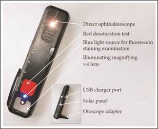
Fig. 1: Arc Light.
All patients were
selected using MR number from Health Information Management System (HIMS) Olive
Track through purposive sampling. First of all, patients were examined by the
Optometrist of the project team and the finding of the selected patients were
noted using Arc light and Ophthalmoscope on the prescribed format and also
entered in the HIMS. Then all selected patients were referred to Medical
Officers (MO’s) on the same day for diagnosis by using Arc light and
Ophthalmoscope. Findings of both were kept separate and they did not know about
each other’s findings. These selected patients were later examined by the
visiting consultant ophthalmologist from LGH by using Arc light and
ophthalmoscope for final evaluation and comparison of findings of the
optometrist and medical officer. Consultant Ophthalmologist findings and
Ophthalmoscope assessments were labeled as the Gold Standards in this study.
All these findings were added in HIMS by each person
separately; MO’s, optometrist and Consultants. This data was also added in SPSS
version 20 by data manger. After that it was further analyzed by Consultant
Researcher according to guidelines to produce an evidence based study.
Sensitivity, specificity, positive predictive value (PPV), negative predictive
value (NPV) and level of agreement were performed on this collected data. This
analysis was used to make a decision about the efficiency of arc light in
comparison with ophthalmoscope and also to evaluate the MO’s training impact.
RESULTS
Results showed that the short term
training of medical officers had only some impact on their skills for making a
correct diagnosis using an arc light or an ophthalmoscope. But Optometrist
produced exceptionally good results and matched with Consultant findings; Gold
standard in this study. Afterwards the validity of arc light was assessed using
sensitivity and specificity analysis. Findings showed that arc light produced
excellent results or almost in parallel to Ophthalmoscope, another Gold
standard tool, if it was used by Optometrist or Consultant Ophthalmologist.
Results of the right eye when observed through Arc light and
Ophthalmoscope by Medical Officers and Consultant showed that there were 23
patients who were classified by Consultant as DR positive cases by using Arc
light while only 11 patients out of 23 were rightly classified by Medical
officers, Table 1. Remaining 12 subjects were misclassified into DR Negative
and others. Similarly, 19 subjects were DR negative or diagnosed as normal by Consultant.
Here only 2 cases out of 19 were wrongly classified into other categories. In
the category of people having other diseases were rightly classified by MO.
Chi-square test of association showed a strong relationship between these two
types of observations. Further Kappa test had a value of 0.506 which showed
that there was 50% level of agreement between Consultant and MO findings about
Right eye through Arc Light.
Similarly, Optometrist findings were also compared with
Consultant; Gold standard in this study. Their findings 100% matched with the
consultant findings in case of RE diagnosis through Arc light, Table 2.
After observing right eye, Arc Light was used to assess the
problems of LE by both; MO and Consultant. Almost similar findings were recorded
for left eye. Here association results were also significant. Kappa test value
shows that there was 54.9% agreement in both observers. Medical Officers (MO)
were not good at diagnosing DR positive cases of Left eye as 10 out of 17 were
wrongly classified, Table 3.
again 100% performance by Optometrist. Their findings matched 100%
with the consultant findings, table 8.
The most important part of the study was
to validate the Arc light as an efficient tool for diagnosis of DR cases and
others. For this purpose, consultant findings on both, Arc light and
Ophthalmoscope were compared and matched. Cross table and Bar chart analysis
highlighted that both results matched 100%. It shows that Arc light can be an
effective tool for diagnosis, table 8.
Now, same procedure was performed for Left eye by Consultant. In this case only one case out of 46 subjects
was misclassified through Arc light. Performance analysis shows that there was
96.6% level of matching in the consultant findings through two different tools,
table 9.
Validity
analysis of the Arc light was done and compared its findings with
Ophthalmoscope. In this case only DR positive and DR Negative cases of RE were
compared through both diagnosing tools. Arc light produced 100% sensitivity,
specificity, PPV, NPV and accuracy. In addition to these values, Confidence
intervals were also given to see the range of accuracy and measurements, table
10.
Table 11: Validation
parameters of RE.
|
Statistic
|
Formula
|
Value
|
95% CI
|
|
Sensitivity
|

|
100.00%
|
85.18% to 100.00%
|
|
Specificity
|

|
100.00 %
|
82.35% to 100.00%
|
|
Disease prevalence
|

|
54.76% (*)
|
38.67% to 70.15%
|
|
Positive Predictive Value
|

|
100.00% (*)
|
|
|
Negative Predictive Value
|

|
100.00% (*)
|
|
|
Accuracy
|

|
100.00% (*)
|
91.59% to 100.00%
|
These
two tools were also applied on LE diagnosis by consultant. But when we
validated the Arc light findings with Ophthalmoscope for DR positive and DR
negative cases, one case was misdiagnosed by Arc light. So, here sensitivity,
NPV and Accuracy reduced to 94.4%, 95.24% and 97.37% from 100% respectively,
table 12 and 13.
Table
12: Validation parameters of LE.
|
Statistic
|
Formula
|
Value
|
95% CI
|
|
Sensitivity
|

|
94.44%
|
72.71% to 99.86%
|
|
Specificity
|

|
100.00 %
|
83.16% to 100.00%
|
|
Disease prevalence
|

|
47.37% (*)
|
30.98% to 64.18%
|
|
Positive Predictive Value
|
|
100.00% (*)
|
|
|
Negative Predictive Value
|

|
95.24% (*)
|
74.86% to 99.26%
|
|
Accuracy
|

|
97.37% (*)
|
86.19% to 99.93%
|
DISCUSSION
The Arc light ophthalmoscope is emerging as a reliable, low-cost
alternative to the standard direct ophthalmoscope. The cost of an Arclight
ophthalmoscope is significantly lower than a direct ophthalmoscope or
comparable instruments. Comparing the current price of Heine direct
ophthalmoscope (USD $365), one can buy 48 Arclight ophthalmoscopes at their
marketed bulk order price (USD $7.5). Arclight is the only direct
ophthalmoscope that is specifically designed for low-income settings15.
However, it would be useful in medical training and education across the globe
by providing an affordable direct ophthalmoscope for medical students. In
comparison to other low-cost direct ophthalmoscopes16,17 the
Arclight has an adjustable lens power with three power settings (+4, −3, and −6 diopters). These lenses will be sufficient for most of the
patient and examiner refractive error. Arc Light also has an additional
attachable otoscope which is helpful to examine ear problems (Figure 1).
We found that findings of
medical officers for right eye and left eye using Arc Light had 50% and 54.9%
agreement respectively with findings of Consultant who was gold standard in
this study and more technical person in eye care. When the medical officer used
the Ophthalmoscope for the assessment of same case’s RE and LE, they got 61%
and 62% agreement with consultant findings. In this study, more than one
medical officer was involved and got training on both tools. So it was planned
to see the individual findings and their agreement with consultant findings.
When split analysis was performed it was observed that there was huge element
of heterogeneity among MO’s in the performance and accuracy. This detailed
analysis showed that they were not at same level and Kappa test also reported
23.4% to 75% level of agreement for RE through ARC Light. For LE it was 39.4%
to 76.5%. In both cases; RE and LE through arc light, MO’s accuracy was 31.8%
to 100% in different doctors when they used Ophthalmoscope for RE and similarly
45.5% to 100% for LE. Overall it can be seen that, MO’s mostly got confused and
gave wrong assessment when they diagnosed those patients who have DR positive
status. In the case of DR negative and others diseases their accuracy was
comparatively good. Lowe et al18 in a similar study, in which
examination was performed by final-year medical students, found no clinically
significant difference between the Arclight ophthalmoscope and the Heine K180
direct ophthalmoscope in terms of accuracy of the vertical cup to disc ration
(VCDR) measurement and with a similar proportion of examinations yielding a ≥ 0.2 difference in the VCDR compared to the reference standard for
both the Arclight and Heine ophthalmoscopes. Importantly, 85% of Arclight
examinations yielded VCDR estimation, compared to 61% with the Heine
ophthalmoscope. Medical-students found that the Arclight was much easier to use
than Heine ophthalmoscope. Moreover the study also found that the LED bulb used
in the Arclight ophthalmoscope was better tolerated by the subjects during ocular
examination, with considerably lower scores for both “glare” and “length of
examination”.
Arc light ophthalmoscope has a solar powered battery which makes
it useful even in remote, rural areas with interrupted power supply and also
cuts the cost of buying new batteries regularly. Our study assessed the
accuracy of the Arc light ophthalmoscope in detecting pathologies in the retina
and it could be used to detect diabetic retinopathy. With such a low cost, Arc
light has the capacity to be much more widely available and will improve training
opportunities and examination of the diabetic retinopathy by medical
specialists in rural areas. Earlier
detection and management of retinopathy will improve the prognosis of the
patients with a less likelihood of progression to blindness.
Blundell R et al19 compared Arclight with traditional
direct ophthalmoscope to examine retinal diseases and found that Arclight was
equally effective in terms of identification of clinical signs and making
correct diagnosis and observers found more ease in using Arclight. Arc light
could be helpful in better training of fundoscopy and easy access to direct
ophthalmoscopes in low budget settings.
In another study by McComiskie et al20 Panoptic versus
conventional direct ophthalmoscope was compared in a group of ‘naïve’ first
year medical students to determine which would be more suitable for
non-ophthalmoligists. Their results showed that the medical students found the
panoptic (PO) much easier to use, with accuracy of rating the VCDR similar to
the conventional direct ophthalmoscope.
We also compared the findings of
Optometrist with gold standard; Consultant. They had excellent accuracy and
level of agreement with the findings of the consultant. Only one case was
misdiagnosed by Optometrist out of 184 cases.
Comparison was also made between
findings of the Consultant with Arc light and Ophthalmoscope. As a whole, only
one case was misdiagnosed through Arc light. Validity of Arc Light versus
Ophthalmoscope was evaluated using sensitivity and specificity analysis.
Overall the Arc Light showed good results in its validity test. Sensitivity and
specificity of Arc light were 100% in RE. But in LE its sensitivity reduced was
to 94.4%. Overall Arc light produced excellent results. The Ophthalmoscope was
used as a Gold standard versus the Arc Light in this analysis.
The limitation of the study was that it was done at
two centres. We have planned to extend this study to other centers to increase
the number of patients examined. Moreover medical students will also be
included in the study to get another perspective.
CONCLUSION
We found that Medical officers had some difficulty
in diagnosing DR positive cases with Arc light. While Optometrists were better
at diagnosis and using these two tools. Furthermore Arc Light is nearly as
efficient tool as an Ophthalmoscope when used by the consultants. Arc light is easy to use and provides comparable results when
examining diabetic retinopathy. It is capable of improving easy access to
equipment in low-budget setups around the world and improvingfundoscopy skills
in eye care workers and diagnosis of retinal diseases. On the basis of these findings we recommend that it is important to train
the MO’s before asking them to use the Arc light. As MO’s are not proficient in
eye care, therefore it is better to introduce Optometrists along with medical
officers at BHU level on permanent basis where they can work in outpatients
department. Arc light can be used as a replacement of Ophthalmoscope for
diagnosing DR or other diseases as shown by the sensitivity and specificity
analysis in this study.
Author’s
Affiliation
Prof.
Muhammad Moin
Head of Department of Ophthalmology
PGMI/LGH/AMC, Lahore
Dr.
Asif Manzoor
Vitreoretinal Fellow,
Lahore General Hospital,
Lahore.
Dr.
Farah Riaz
Project Manager
Fred Hollows Foundation
Islamabad
Role of
Authors
Prof.
Muhammad Moin
Study Design, Manuscript writing and Critical review
Dr.
Asif Manzoor
Study Design, Data Collection and Critical review
Dr.
Farah Riaz
Manuscript writing, Data analysis, Study design
Conflict of Interests
None.
ACKNOWLEDGEMENTS
The study was supported by
unrestricted educational grant by the Fred Hollows Foundation.
REFERENCES
1. Dineen B, Bourne RR, Jadoon Z, Shah SP, Khan MA, Foster A, Gilbert CE,
Khan MD. Causes of blindness and visual impairment in Pakistan. The
Pakistan National Blindness and visual impairment survey; Pakistan National Eye
Survey Study Group.Br J Ophthalmol. 2007 Aug; 91 (8): 1005-10.
2. Sherin A. National diabetes action plan of Pakistan: Need and
challenges. Khyber Med Univ J. 2015; 7 (1): 1-2.
3. Basit A, Fawwad A, Baqa K.
Pakistan and diabetes - a country on the edge. Diabetes Res Clin Pract. 2018
Nov 10. pii: S0168-8227(18)31639-5.
4. Shaw JE, Sicree RA, Zimmet PZ. “Global estimates of the prevalence
of diabetes for 2010 and 2030,” Diabetes Research and Clinical Practice,
2010; Vol. 87, No. 1: pp. 4–14.
5. Sivaprasad S, Gupta B, Gulliford MC et al. Ethnic variations in the
prevalence of diabetic retinopathy in people with diabetes attending screening
in the United Kingdom (DRIVE UK), PLoS ONE, vol. 7, no. 3, Article ID e32182,
2012.
6. Klein BEK. “Overview of epidemiologic studies of diabetic
retinopathy,” Ophthalmic Epidemiology, 2007; Vol. 14, No. 4: pp. 179–
183.
7. Ting DS, Cheung GC, Wong TY. Diabetic
retinopathy: global prevalence, major risk factors, screening practices and
public health challenges: a review. Clin Exp Ophthalmol. 2016 May; 44
(4): 260-77.
8. Tham, Y.C., et al. Global prevalence of glaucoma and projections of
glaucoma burden through 2040: a systematic review and meta-analysis.
Ophthalmology, 2014; 121 (11): p. 2081-90.
9. Heijl A, Leske MC, Bengtsson B, Hyman L, Bengtsson B, Hussein M.
“Reduction of intraocular pressure and glaucoma progression: results from the
Early Manifest Glaucoma Trial,” Archives of Ophthalmology, 2002; Vol.
120, No. 10: pp. 1268–1279.
10. The Diabetes Control
and Complications Trial Research Group, “The effect of intensive treatment of
diabetes on the development and progression of long-term complications in
insulin dependent diabetes mellitus,” The New England Journal of Medicine,
1993; Vol. 329, No. 14: pp. 977–986.
11. Bastawrous A, Hennig BD. “The global inverse care law: a distorted
map of blindness,” British Journal of Ophthalmology, 2012; Vol. 96, No.
10: pp. 1357–1358.
12. Das T, Ackland P, Correia M, et al. Is the 2015 eye care service
delivery profile in Southeast Asia closer to universal eye health need! Int Ophthalmol.
Doi 10.1007/s10792-017-0481-y
13. Palmer JJ, Chinanayi F, Gilbert A et al. “Mapping human resources
for eye health in 21 countries of sub-Saharan Africa: current progress towards
VISION 2020,” Human Resources for Health, 2014; Vol. 12, No. 1: article
44.
14. Resnikoff S,
Felch W,
Gauthier TM,
Spivey B. The number of
ophthalmologists in practice and training worldwide: a growing gap despite more
than 200,000 practitioners. Br J Ophthalmol. 2012 Jun;
96 (6): 783-7.
15. Blaikie
A, Sandford-Smith J, Tuteja SY, Williams CD, O'Callaghan C. Arclight: a pocket ophthalmoscope for the 21st century.BMJ. 2016 Dec. 14; 355: i6637.
16. Armour
RH. Manufacture and use of homemade ophthalmoscopes: a 150th anniversary
tribute to Helmholtz, British Medical Journal, 2000; Vol. 321, No. 7276:
pp. 1557–1559.
17. Mandal
N, Harborne P, Bradley S et al. “Comparison of two ophthalmoscopes for
direct ophthalmoscopy,” Clinical and Experimental Ophthalmology, 2011;
Vol. 39,
No. 1: pp. 30–36.
18. Lowe J, Cleland RC, Mgaya E, Furahini G,
Gilbert CE, Burton MJ, Philippin H.
The Arclight Ophthalmoscope: A Reliable Low-Cost Alternative to the Standard
Direct Ophthalmoscope; Journal of Ophthalmology Volume 2015: 1-6.
19. Blundell
R, Roberts D, Fioratou E et al. Comparative
evaluation of a novel solar powered low-cost ophthalmoscope (Arclight) by eye
healthcare workers in Malawi. BMJ Innov. 2018 Apr; 4 (2): 98–102.
20. McComiskie
JE, Greer RM, Gole GA. “Panoptic versus conventional ophthalmoscope,” Clinical
& Experimental Ophthalmology, 2004; Vol. 32, No. 3, pp. 238–242.